
![]()
OMRF Quantitative Analysis Core
HiLIFE University of Helsinki

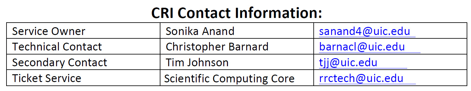
UIC Scientific Computing Core
Shared Instrument-UMN Hormel Institute


VU VIDL

UIC Preclinical Imaging Core
VUMC Storeroom


Monash MCEM and DoCE
TAMU Materials and Pavement
![]() Access to Lab anLAB AGREEMENT for LIMS (1)d Equipment (1)
Access to Lab anLAB AGREEMENT for LIMS (1)d Equipment (1)